Pigmentation Clinic Singapore: Understanding Deep Dermal Pigmentation vs Surface Pigmentation
Many people assume that all pigmentation can be treated in the same way. However, pigmentation can develop at different depths within the skin. Some forms affect the skin’s surface layers, while others originate deeper within the dermis. Understanding the difference between deep dermal pigmentation and surface pigmentation is important because the location of the pigment often influences how it appears and how it may be managed. At a pigmentation clinic Singapore patients often discover that seemingly similar dark spots may have very different underlying causes and depths within the skin.
A proper assessment at a pigmentation clinic can help determine the type of pigmentation present and guide treatment planning. If you are considering visiting a pigmentation clinic Singapore, learning about the distinction between surface and deep pigmentation can help you better understand your condition and the treatment options that may be recommended.
What Is Surface Pigmentation?
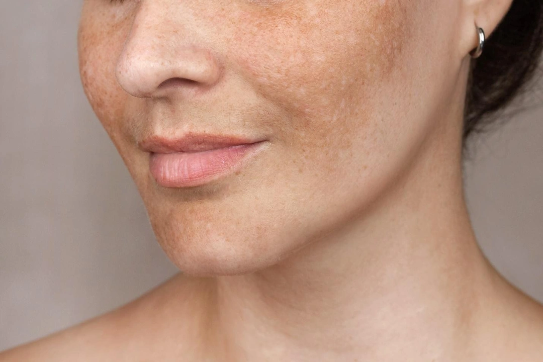
Surface pigmentation, also known as epidermal pigmentation, occurs when excess melanin accumulates within the outermost layers of the skin. Because the pigment is located closer to the skin’s surface, these lesions often appear light to dark brown in colour and may have relatively distinct borders. Common examples of surface pigmentation include freckles, solar lentigines (sunspots), and certain forms of post-inflammatory hyperpigmentation that develop after acne, eczema, or minor skin injuries.
Ultraviolet (UV) exposure is a major contributing factor, as it stimulates melanin production and can cause existing pigmentation to become more noticeable over time. In many cases, epidermal pigmentation may respond differently to treatment than deeper forms of pigmentation because the pigment is more accessible within the skin’s upper layers.
What Is Deep Dermal Pigmentation?
Deep dermal pigmentation occurs when melanin or pigment-producing cells are located within the deeper dermal layers of the skin rather than the epidermis. Because the pigment lies beneath the surface, it often appears grey, blue-grey, or slate-coloured due to the way light is scattered through the skin. Common examples include Hori’s nevus, acquired dermal melanocytosis, and certain congenital pigmentary conditions. Unlike surface pigmentation, dermal pigmentation may not always have sharply defined borders and can sometimes be mistaken for melasma or age-related pigmentation.
This distinction is important because deeper pigment deposits often behave differently and may require a different management approach. In Asian populations, dermal pigmentation is relatively common and can be challenging to identify without a thorough clinical assessment. Understanding whether pigmentation is located within the epidermis or dermis is often one of the first steps in developing an appropriate treatment plan.
How Can You Tell Whether Pigmentation Is Deep or Superficial?
Determining whether pigmentation is located within the epidermis (surface layer) or dermis (deeper layer) is an important step in diagnosis. The depth of pigmentation often affects its colour, appearance, and potential response to treatment. While certain characteristics can provide clues, pigmentation disorders may sometimes overlap, making professional assessment necessary.
Colour and Appearance of the Pigmentation
One of the most noticeable differences between superficial and deep pigmentation is colour. Superficial pigmentation typically appears light brown to dark brown because the excess melanin is located closer to the skin’s surface. Examples include freckles, sunspots, and some forms of post-inflammatory hyperpigmentation.
Deep dermal pigmentation often appears grey, blue-grey, or slate-coloured. This occurs because light is scattered differently when pigment is located deeper within the skin. Conditions such as Hori’s nevus commonly exhibit this characteristic appearance.
Distribution and Location on the Face
The location and pattern of pigmentation can also provide useful diagnostic clues. Superficial pigmentation is often found in sun-exposed areas and may present as isolated spots or patches. Deep dermal pigmentation, on the other hand, frequently develops on areas such as the cheekbones, temples, and around the eyes. However, pigmentation patterns are not always straightforward.
Why a Professional Assessment Is Important
Although colour and location can offer hints about pigment depth, it is not always possible to make an accurate diagnosis through visual inspection alone. Melasma, for example, may contain epidermal, dermal, or mixed pigment components, making it difficult to classify without a detailed evaluation.
During a consultation, a doctor may assess factors such as the pigmentation’s appearance, distribution, duration, progression, and response to previous treatments. Identifying whether the pigmentation is superficial, deep, or mixed is essential for developing an appropriate treatment strategy and setting realistic expectations for results.
Why Does Pigment Depth Matter for Treatment?
The depth of pigmentation plays a significant role in determining the most appropriate treatment approach. Since pigment can be located at different levels within the skin, treatment outcomes may vary depending on whether the pigmentation is superficial, deep, or mixed.
Accurately identifying pigment depth is important because it can affect:
- The type of treatment that may be recommended
- The number of treatment sessions required
- How quickly visible improvements may occur
- The likelihood of pigmentation recurring
- The overall expectations for treatment outcomes
Can a Person Have Both Deep and Surface Pigmentation?
Yes. It is possible for an individual to have more than one type of pigmentation at the same time. In fact, mixed pigmentation is relatively common, particularly among individuals with Asian skin.
Examples of combined pigmentation conditions include:
- Melasma occurring alongside sunspots (solar lentigines)
- Hori’s nevus coexisting with melasma
- Post-inflammatory hyperpigmentation developing in areas affected by other pigmentation disorders
When multiple pigmentary conditions are present, diagnosis can become more challenging because different types of pigmentation may overlap or appear similar.
When Should You Visit a Pigmentation Clinic?
While some forms of pigmentation may fade gradually over time, others can persist, worsen, or recur despite using skincare products. Seeking professional assessment may be beneficial if you notice any of the following:
- Pigmentation that does not improve with over-the-counter products
- Dark patches that continue to spread or become more noticeable
- Pigmentation that repeatedly returns after previous treatments
- Uncertainty about the type or cause of your pigmentation
- Multiple forms of pigmentation appearing on the face
Conclusion
Understanding whether pigmentation is located on the skin’s surface or within the deeper dermal layers is an important step in achieving an accurate diagnosis and appropriate treatment plan. Because different types of pigmentation can appear similar, a professional assessment can help identify the underlying condition and determine the most suitable approach for your skin concerns.
If you are concerned about persistent pigmentation, melasma, Hori’s nevus, or other forms of facial pigmentation, contact us, at:
One Face Clinic – Pigmentation | Melasma Treatment | Acne Singapore
1 Tras Link, #02-01 Orchid Hotel Singapore, Singapore 078867 | +65 6222 2262